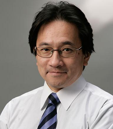
PL-1 Molecular mechanism of CRISPR and structure-based development of genome editing tool towards medical applications
Osamu Nureki
Department of Biological Sciences, Graduate School of Science,
The University of Tokyo, 2-11-16 Yayoi, Bunkyo-ku, Tokyo 113-0032, Japan
The CRISPR-associated endonuclease Cas9 can be targeted to specific genomic loci by single guide RNAs (sgRNAs). We solved the crystal structure of Cas9, from 4 sources (984 a.a. to 1,629 a.a.), complexed with sgRNA and its target DNA at atomic resolutions. These high-resolution structures combined with functional analyses revealed the generality and diversity of molecular mechanism of RNA-guided DNA targeting by Cas9, and uncovered the distinct mechanisms of PAM recognition. On the basis of the structures, we succeeded in changing the specificity of PAM recognition, which paves the way for rational design of new, versatile genome-editing technologies. Recently, our high-speed atomic force microscopy (HS-AFM) analysis of SpCas9 visualized real-space and real-time dynamics of Cas9. We further solved the crystal structure of type-V CRISPR, Cpf1 in complex with crRNA and target dsDNA. The structure explains striking similarity and major differences between Cas9 and Cpf1.
References- “Crystal Structure of Cas9 in Complex with Guide RNA and Target DNA” H. Nishimasu, F. A. Ran, P. D. Hsu, S. Konermann, S. I. Shehata, N. Dohmae, R. Ishitani, F. Zhang F and O. Nureki.
Cell 156, 935-949 (2014). doi: 10.1016/j.cell.2014.02.001. - “Crystal structure of Staphylococcus aureus Cas9” H. Nishimasu, L. Cong, W. X. Yan, F. A. Ran, B. Zetsche, Y. Li, A. Kurabayashi, R. Ishitani, F. Zhang and O. Nureki
Cell 162, 1113-1126 (2015). doi: 10.1016/j.cell.2015.08.007. - “Structure and Engineering of Francisella novicida Cas9” H. Hirano, J. S. Gootenberg, T. Horii, O. O. Abudayyeh, M. Kimura, P. D. Hsu, T. Nakane, R. Ishitani, I. Hatada, F. Zhang, H. Nishimasu and O. Nureki.
Cell 164, 950-961 (2016) doi: 10.1016/j.cell. - “Structural basis for the altered Pam specificities of engineered CRISPR-Cas9” S. Hirano, H. Nishimasu, R. Ishitani and O. Nureki.
Mol. Cell 61, 886-894 (2016) doi 10.1016/j.molcel.2016.02.018. - “Crystal structure of Cpf1 in complex with guide RNA and target DNA” T. Yamano, H. Nishimasu, B. Zetsche, H. Hirano, I. M. Slaymaker, Y Li, I. Fedorova, T. Nakane, K. S. Makarova, E. V. Koonin, R. Ishitani, F. Zhang and O. Nureki.
Cell 165, 949-962 (2016) doi: 10.1016/j.cell.2016.04.003. - “Crystal Structure of the Minimal Cas9 from Campylobacter jejuni Reveals the Molecular Diversity in the CRISPR-Cas9 Systems” M. Yamada, Y. Watanabe, J. S. Gootenberg, H. Hirano, F. A. Ran, T. Nakane, R. Ishitani, F. Zhang, H. Nishimasu and O. Nureki.
Mol. Cell. 65, 1109-1121 (2017). - “Real-space and real-time dynamics of CRISPR-Cas9 visualized by high-speed atomic force microscopy” M. Shibata, H. Nishimasu, N. Kodera, S. Hirano, T. Ando, T. Uchihashi and O. Nureki
Nat. Commun. 8, 1430 (2017).

PL-2 From the discovery of Matrigel and it uses in cell and cancer biology to the identification of thymosin b4 for tissue repair and regeneration
Hynda K. Kleinman
NIH Guest Researcher, Adjunct Professor,
The George Washington University School of Medicine, USA
Basement membranes are thin extracellular matrices that underlie epithelial and endothelial cells and surround smooth muscle, peripheral nerves, and fat cells. Basement membrane is the first matrix to be formed in the developing embryo and has important activity with normal, tumor, and stem cells. A murine tumor matrix extract, termed Matrigel, has provided an abundant source of basement membrane proteins (laminin, collagen IV, heparan sulfate, etc) and a basement membrane mixture that gels at room temperature into a structure similar to the authentic matrix. In vitro, Matrigel promotes the differentiation of many cell types, tissue explants, and stem cells, such as capillary formation by endothelial cells, myelin production by spinal ganglia, milk production by breast epithelial cells, acinar formation by embryonic salivary glands, organoid formation, etc. Matrigel has been used in various in vitro assays for angiogenesis, cell invasion, spheroid formation, organoid formation from a single cell, etc. In vivo, Matrigel has been used to improve/promote tumor xenograft growth, measure angiogenesis, improve heart and spinal cord repair, increase tissue transplant take, etc. Endothelial cells form caplillary-like structures with a lumen by 12 hours when plated on Matrigel. The gene for thymosin beta 4 was induced at 4 hours after plating the endothelial cells on Matrigel, and when the thymosin beta 4 protein was added exogenously to the culture, differentiation was accelerated. Thymosin beta 4, a small 43 Kd protein present in all body fluids and cells, has multiple biological activities, including reducing inflammation, apoptosis, and cytotoxicity while increasing cell migration, stem cell recruitment and differentiation, and tissue repair. Various active sites on thymosin beta 4 and a receptor (ATP synthase) have been identified. Thymosin beta 4 was subsequently found to promote angiogenesis in vivo and to improve dermal and ocular healing in experimental models of injury. It has efficacy in animal models of traumatic brain injury, stroke, multiple sclerosis, heart attack, peripheral neuropathy, liver and kidney fibrosis, hair growth, etc. Furthermore, low thymosin beta 4 levels in the blood correlate positively with disease progression/death vs higher levels which are associated with disease improvement/longer survival. Phase 2 and in some cases phase 3 clinical trials demonstrated its efficacy for both stasis and pressure ulcers and for dry eye and other ocular diseases.
